 English
 عربى
عربى Español
 русский
 中文简体
中文简体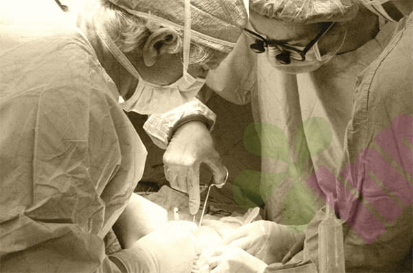


A fracture is much more than just a broken bone. When the external force far exceeds the bone's bearing limit, causing its continuity or integrity to be interrupted, we call it a fracture. Imagine the sudden collapse of the main beam of a bridge - bones are the support of the human body. Once they break, not only will the supporting function be lost, but the surrounding blood vessels, nerves, and muscles are often affected. Especially in open fractures, the tip of the broken bone pierces the skin, forming a wound that is connected to the outside world. This open "door" allows bacteria to enter directly, instantly upgrading a simple bone injury to a high-risk infection event.
Why are open fractures particularly dangerous?
Different from closed fractures with intact skin, the fatal threat of open fractures lies in the exposed wound surface. Based on the size of the wound, the depth of contamination and the degree of soft tissue damage (usually Gustilo classification), doctors will classify it into three degrees: mild (I degree) may be just a small hole punctured by the bone tip; moderate (II degree) accompanied by obvious soft tissue tearing; and severe (III degree) means extensive tissue damage, rupture of blood vessels and nerves, and even severe contamination caused by battlefields or agricultural machinery. The deeper the degree, the risk of infection and disability rises exponentially.
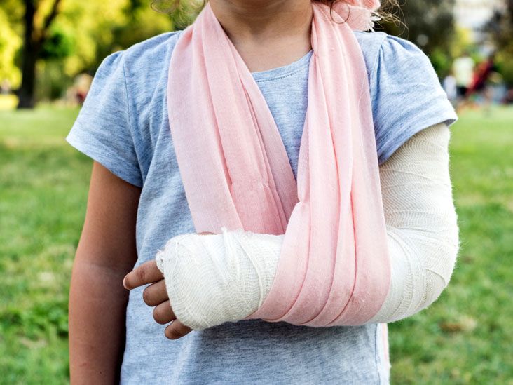
What distress signals will the injured area send out?
The injured often break out in cold sweats due to the severe pain, and any movement of the affected limb will trigger a heart-wrenching cry of pain. Visible limb deformities, abnormal bending angles, or joint movements that should not occur (medically known as "abnormal movements") are all red alerts for fractures. A light touch may feel bone friction - that is the creepy sign of the friction between the broken ends. Open wounds are more direct warnings, and bleeding or directly exposed bone stumps are shocking. These local manifestations are like beacons of the body, urgently announcing the disaster.
What alarms will sound throughout your body?
Don't just focus on the wound! When the blood loss exceeds 500ml (especially pelvic or femoral fractures), the victim may turn pale, have a weak and rapid pulse, and breathe rapidly - this is a precursor to shock. What's more dangerous is that bacteria are proliferating wildly through the wound, which may cause systemic infection within a few hours, manifested as high fever, chills, and confusion. If the limbs are in constant pain, pale, and you can't feel the pulse? Beware of compartment syndrome! This sudden increase in pressure caused by internal bleeding can permanently destroy nerves and muscles in a short period of time.
On-site first aid procedures?
1. Stop bleeding: Immediately cover the wound with a sterile dressing or clean cloth and continue to apply strong pressure. If the arterial bleeding does not stop, the proximal tourniquet is the last resort (record the use time! Loosen it once every hour).
2. Wound protection: Do not flush or stuff the exposed bone back! Cover it tightly with sterile gauze/plastic wrap to prevent contamination.
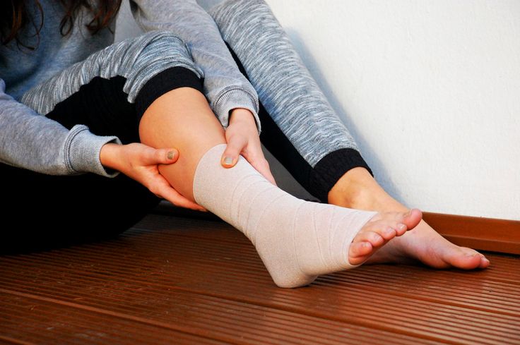
3. Temporary fixation is a key step: use splints, cardboard, or even healthy limbs to fix the upper and lower ends of the fracture above the joint. Remember: it is better to maintain the deformity than to reduce it violently!
4. Anti-shock and rapid transport: Keep warm, lie flat with lower limbs elevated, and quickly rehydrate when conditions permit. Transport to a hospital with debridement capabilities as quickly as possible, and continuously assess vital signs during the journey.
Open fracture debridement?
1. Thorough scrubbing: After anesthesia, use a sterile brush dipped in iodine solution to repeatedly scrub the skin around the wound, and use normal saline to vigorously flush the inside of the wound cavity to wash away pollutants such as mud, debris, etc.
2. Precise excision: Dissect layer by layer like an archaeologist, and completely remove all inactive and contaminated muscles (dark purple in color, no contraction), fascia, and subcutaneous tissue. Treat bones with caution, and only remove completely free small bone fragments.
3. Pulse irrigation: High-pressure pulse water flow deeply rinses every corner of the wound again to dilute residual bacteria.
4. Emergency repair of blood vessels and nerves: If major blood vessels or nerves are found to be broken, they must be immediately anastomosed under a microscope.
5. Stabilize the fracture ends: Select an external fixator (preferred when there is severe contamination) or internal fixation (light contamination and good soft tissue conditions) according to the type of injury.
6. Delayed wound closure: In most cases, the wound is not sutured directly, but covered with negative pressure suction by VSD. After 3-5 days, it is confirmed that there is no infection before closure or flap transplantation.
Broad-spectrum antibiotics are given intravenously immediately after surgery, covering both gram-positive and gram-negative bacteria. Tetanus antitoxin is essential. The affected limb is kept elevated to reduce swelling, and the rehabilitation therapist guides muscle isometric contraction training. Regular X-rays are performed to observe bone healing, and be alert to the potential danger of osteomyelitis - it may lurk for several weeks and then suddenly attack with redness, swelling, heat, and pain. For more information on Innomed®Elastic Bandage Self-Adhesive, refer to the Previous Articles. If you have customized needs, you are welcome to contact us; You Wholeheartedly. At longterm medical, we transform this data by Innovating and Developing Products that Make Life easier for those who need loving care.
Editor: kiki Jia


