
 English
 عربى
عربى Español
 русский
 中文简体
中文简体






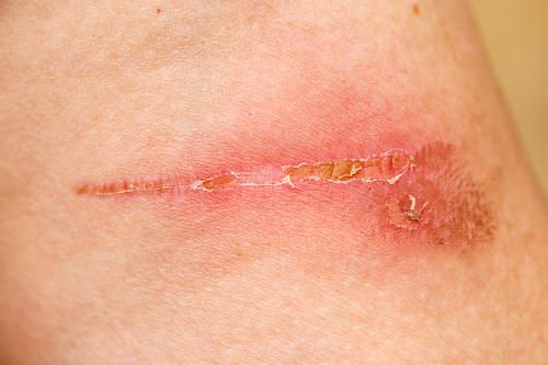
A red ring around a wound is one of the most anxiety-inducing aspects of home wound care. The conditioned reflex of associating red with inflammation, leading to the application of antibiotic ointment, often interferes with the normal healing process. In reality, wound redness can be categorized into at least four distinct types: physiological inflammatory response, contact dermatitis, maceration-associated erythema, and infectious cellulitis, each requiring drastically different treatment approaches.
What does redness look like during a normal healing process?
The inflammatory phase of wound healing is a necessary stage. Within one to three days after injury, the wound and surrounding tissues release vasoactive substances such as histamine, prostaglandins, and leukotrienes. Local capillaries dilate and become congested, manifesting as a pale red or pink halo around the wound edge. The color is uniform and without a clear distinction between light and dark areas. The redness temporarily fades when lightly pressed with a finger and slowly returns upon release. This red halo is usually limited to within three to five millimeters outward from the wound edge, with blurred borders that gradually transition to normal skin color. Simultaneously, bright red granular granulation tissue is visible at the wound base, with moderate and clear exudate. This physiological redness is not accompanied by increased pain, significant increase in local skin temperature, or skin edema beyond the red area. This physiological inflammatory reaction does not require special treatment; regular dressing changes and wound protection are sufficient. As the wound enters the proliferative phase, the redness will gradually subside within a few days.
Which types of redness are caused by external stimuli rather than infection?
One type of redness that is easily misdiagnosed as infection is actually caused by chemical irritation or allergic reactions to the skin around the wound from dressing adhesives, disinfectants, or skin care product ingredients. This type of redness is characterized by its area and shape closely matching the contact substance; for example, a rectangular red patch perfectly matching the shape of the tape, or a clearly defined red mark appearing in the area after iodine disinfection. The redness is usually brighter than infectious erythema, sometimes accompanied by small papules or even small blisters. Itching is significant, but pain is mild, and there are no signs of infection in the wound's base or exudate. The treatment involves discontinuing the suspected irritant and using silicone-adhesive dressings and colorless, non-irritating skin protectants. The redness usually begins to subside on its own within 48 hours after the irritant is removed. Another type of redness related to moisture is caused by wound exudate soaking the surrounding skin. The skin around the wound becomes white and swollen due to prolonged contact with exudate or sweat. After removing the dressing and exposing it to air, it turns from white to red, with a moist, smooth skin surface and even superficial erosion. The key to treating maceration erythema is exudate management, which involves switching to dressings with greater absorbency or water-locking properties, and applying a skin protective film or zinc oxide ointment around the wound to create an isolation barrier.
What are the characteristics of infectious erythema that warrant high vigilance?
Localized, spreading erythema caused by infection, clinically known as cellulitis, is the only type of erythema among the four types requiring emergency medical intervention. Several key characteristics help identify it. In terms of color, infectious erythema is usually dark red or deep red, not pink, and the color is uneven, with the central area being the darkest and gradually fading towards the edges. Regarding the borders, the biggest difference from normally healing erythema is the indistinct borders; there is no clear boundary between the red area and normal skin. Pressing on the red area for a few seconds and then releasing it will not significantly lighten the redness. This pressure test is somewhat difficult to interpret on darker skin, but it still has reference value. In terms of extent, infectious erythema continuously expands outwards; the red area can be visibly increased within 24 hours. The rate of expansion is the most important dynamic indicator for differentiating infection from normal inflammation. Accompanying signs include a significantly higher skin temperature in the erythematous area compared to the contralateral normal skin, a swollen and firm feel upon touch rather than simple surface redness, and persistent pain even at rest, sometimes presenting as throbbing pain. The appearance of systemic reactions such as fever, chills, and swollen and tender lymph nodes on the same side indicates that the infection has spread beyond the local area, requiring immediate medical attention and intravenous antibiotics. It is particularly important to emphasize that infections in diabetic patients and those with lower extremity arterial ischemia often spread faster than in the general population. Pain may be less pronounced due to neuropathy. These high-risk individuals should lower their risk threshold for medical attention if any redness around the wound exceeds the normal range, and should not wait for it to subside on its own at home. For more information on Innomed® Polyurethane foam dressing, refer to the Previous Articles. If you have customized needs, you are welcome to contact us; You Wholeheartedly. At long-term medical, we transform this data by innovating and developing products that make life easier for those who need loving care.
Editor: kiki Jia


