 English
 عربى
عربى Español
 русский
 中文简体
中文简体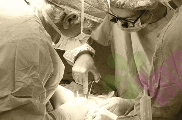


People who have undergone abdominal surgery sometimes find a pale yellow or clear fluid with oily droplets seeping from the incision when removing sutures or changing dressings. There is no redness, pain, or fever, but the gauze is always damp. This is very likely fat liquefaction. Fat liquefaction is a specific problem during the surgical incision healing process, more common in patients with thicker abdominal fat, and also frequently seen after surgeries using electrocautery or high-frequency electrocoagulation instruments. It is not an infection, but improper treatment can easily lead to secondary infection.
What is fat liquefaction, and how is it different from infection?
In simple terms, fat liquefaction occurs when fat cells beneath a surgical incision rupture and break down due to ischemia, thermal damage, or mechanical stimulation, releasing neutral fat as free oil droplets. These droplets mix with tissue exudate and flow out from the incision. Typical fat liquefaction exudate is a clear, pale yellow or yellowish liquid; when collected with gauze, it leaves a noticeable oily residue, and after drying, a ring of grease remains on the gauze. The exudate has no odor, the skin around the incision is not red or only slightly red, and the patient does not experience systemic discomfort. This is completely different from surgical site infection. Infected exudate is cloudy, yellowish-white or yellowish-green, with a fishy or fecal odor; the incision edges are noticeably red and swollen, the skin temperature is elevated, and there is distinct pain upon pressure; in severe cases, the patient may develop a fever and elevated white blood cell count. Distinguishing between these two is crucial because the treatment approaches are completely opposite. Fat liquefaction requires maintaining unobstructed drainage and promoting granulation tissue growth, while infection requires thorough debridement and the use of antibacterial dressings.
Who is most prone to fat liquefaction?
Fat liquefaction doesn't happen to everyone; there are several clearly defined risk factors. The most direct is the thickness of abdominal wall fat; patients with subcutaneous fat exceeding two centimeters have a significantly higher incidence. Adipose tissue itself has a relatively poor blood supply; the thicker the fat layer, the less blood perfusion in the central area, making the ends of the fat flaps on both sides prone to ischemia and necrosis after surgical incision. The use of electrocautery is also a common cause. The heat generated when cutting through the fat layer can reach over 100 degrees Celsius, a temperature that fat cells cannot withstand, causing their cell membranes to rupture instantly. Experienced surgeons will use a low-power electrocautery to quickly penetrate the fat layer or switch to a scalpel to cut the fat, but in some surgical conditions, it is difficult to completely avoid thermal damage. Obese patients, those with diabetes, and those with hypertension generally have poor microcirculation and poor postoperative tissue repair capabilities, increasing their risk of fat liquefaction compared to other patients. Furthermore, the location of the incision also matters; the blood supply differs between the upper and lower abdomen. The lower abdomen often has a thicker fat layer with poorer blood supply, increasing the probability of liquefaction.
What should be done after fat liquefaction occurs?
If fat liquefaction and exudation are found at the incision, the worst thing to do is repeatedly squeeze the incision in an attempt to drain the fluid. Squeezing will force fat debris, which would otherwise be confined to the dead space, into the normal tissue spaces, artificially expanding the area of contamination. The correct approach is to first gently dab the exudate with a sterile cotton swab to observe its characteristics. After confirming that there is no purulent discharge or foul odor, drainage can be performed. If the amount of exudate is small, with only a small damp patch on the gauze, special drainage is not necessary. Simply increase the frequency of dressing changes to once a day or every other day, covering the area with sterile dry gauze to absorb the exudate. At the same time, apply appropriate pressure with an abdominal binder to reduce subcutaneous cavity. Most mild liquefaction will stop on its own within one to two weeks, and the incision will gradually heal.
If the exudate is significant, soaking several gauze pads daily, intervention is necessary. A common method is to place a drainage strip. Using sterile forceps, insert an iodine-soaked gauze strip or Vaseline-soaked gauze strip below the incision or at the deepest point of the fluid cavity, leaving one end of the gauze strip outside the incision to allow liquefied oil and exudate to drain out. The drainage strip should be changed daily. Each time, flush the cavity with saline solution to remove any remaining oil droplets and debris before inserting a new drainage strip. As the exudate gradually decreases, the length of the drainage strip should be gradually shortened until it is completely removed, allowing the incision to close naturally. This process typically takes one to three weeks.
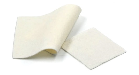
For chronic liquefaction cavities with recurrent exudation lasting more than two weeks, some functional wound dressings can be useful. Alginate dressings are highly absorbent materials made from fibers extracted from brown algae. Upon contact with exudate, they form a soft gel that can fill the liquefied cavities, absorbing oil and exudate while keeping the wound moist and preventing maceration. When using alginate dressings, they need to be cut into strips and inserted into the cavity, then covered with dry gauze for fixation. They can be changed every two days, which is more convenient than changing drainage strips daily, and alginate dressings are less irritating to granulation tissue. Foam dressings can also be used in the superficial stage of liquefaction. If the cavities are already relatively shallow, a polyurethane foam dressing can be directly applied to the incision. It can absorb exudate while providing moderate pressure to help close the cavity. However, foam dressings are not suitable for deep liquefaction cavities because the dressing only applies to the surface and cannot address the problems deep within the cavity.
For more information on Innomed®Alginate Dressing, refer to the Previous Articles. If you have customized needs, you are welcome to contact us; You Wholeheartedly. At long-term medical, we transform this data by innovating and developing products that make life easier for those who need loving care.
Editor: kiki Jia


