
 English
 عربى
عربى Español
 русский
 中文简体
中文简体






The ability to take a photo of a wound with a mobile phone, upload it, and receive assessment suggestions is no longer just a concept in medical applications both domestically and internationally. Many hospital wound clinics and online medical platforms have opened text-based consultation channels, and some specialized wound assessment apps have also entered home care scenarios. However, most people are unclear about the actual capabilities and limitations of this technology, and are prone to either over-trusting AI as a doctor or completely rejecting it, feeling that a personal examination is necessary. Only by objectively understanding the working principles and applicable scope of AI wound assessment can it be used where it is needed.
How does AI deduce wound information from photos?
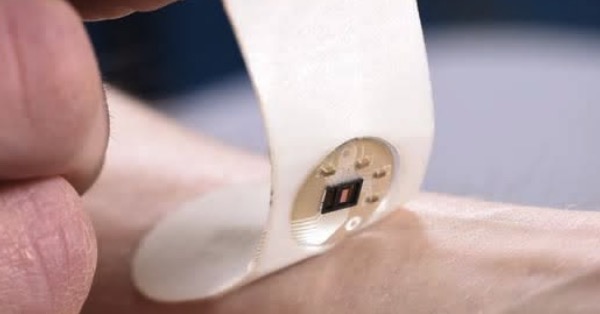
The core technology of AI wound assessment is the segmentation and classification of two-dimensional images using deep learning convolutional neural networks. The algorithm is trained on tens or even hundreds of thousands of wound photographs already annotated by professional wound therapists, learning to identify which pixels in the image belong to the wound area, which are surrounding normal skin, and what different colored tissues within the wound area correspond to. Black areas may be eschar, yellow may be necrotic tissue or fatty tissue, red may be granulation tissue, and white may be maceration or tendon. After segmentation, the AI calculates the actual wound size based on the pixel area of the wound area and the proportion of reference objects in the photo, and then combines this with the color distribution ratio to give the tissue type percentage, such as 60% granulation tissue, 30% necrotic tissue, and 10% eschar. Some more sophisticated algorithms can also identify the integrity of the wound edges, the extent of redness and swelling in the surrounding skin, and the distribution characteristics of exudate on the dressing or wound surface. The entire process, from uploading the photo to generating the report, can be completed within tens of seconds. The output typically includes wound area, tissue type ratio, estimated depth, and a preliminary classification recommending medical attention.
Can AI really determine whether a wound is infected based on just one photo?
The answer to this question needs to be considered in a layered way. AI can, to some extent, identify visual feature combinations that are highly associated with infection, such as a wound color changing from reddish to grayish or yellowish-green, the appearance of large, regular patches of erythema on the surrounding skin, and the purulent, cloudy appearance of exudate in photos. These feature patterns have been repeatedly labeled in the training data, and the algorithm learns the statistical correlation between feature combinations and infection labels. However, under current technological conditions, AI cannot determine the odor of exudate from static images, cannot palpate changes in skin temperature, cannot perceive the nature of the patient's pain, and cannot measure laboratory indicators such as white blood cell count and C-reactive protein. These dimensions are precisely the indispensable basis for clinical diagnosis of wound infection. Therefore, the infection risk warnings given by AI from photos should be understood as a preliminary screening tool rather than a diagnostic conclusion. Its value lies in reminding patients or their families that there are some alarming visual signals about the wound and suggesting that a comprehensive evaluation by professionals be conducted as soon as possible, rather than replacing professionals in determining whether antibiotics or wound cleaning are necessary.
Should AI assessments be trusted?
This is the most common and crucial point of contention when using AI for assessment. AI's judgment logic is based on statistical models; it doesn't tire or miss anything, but it lacks understanding of subjective experiences such as pain and the impact of activity. When AI continuously prompts for medical attention while the patient feels well, it's necessary to examine the specific risk points indicated by the AI. If the risk point points to abnormal tissue type, such as a report showing eschar or necrotic tissue accounting for more than 30%, this means there is a large amount of inactive tissue within the wound, making healing difficult without debridement; even if the wound area is small, medical attention is necessary. If the risk point points to an increase in wound area instead of a decrease, with reports showing no trend of reduction over two consecutive weeks, medical attention should also be considered. If the AI indicates that the surrounding redness and swelling exceeds a certain proportion of the wound area, it may correspond to the early stage of cellulitis, requiring prompt in-person medical attention. Conversely, if the AI risk rating is low, with a rosy wound color, weekly reduction in area, and normal surrounding skin, such wounds can continue to be cared for at home, with regular photo monitoring of the trend. AI's suggestions are probabilistic judgments output by statistical models. The final decision requires a comprehensive consideration of the patient's overall condition, pain perception, and past healing experience. However, in uncertain situations, it's better to make an extra trip to the clinic than to let a wound that could be treated early become chronic. For more information on Innomed® Silver Ion Dressing Foam, refer to the previous articles. If you have customized needs, you are welcome to contact us; You Wholeheartedly. At long-term medical, we transform this data by innovating and developing products that make life easier for those who need loving care.
Editor: kiki Jia


