
 English
 عربى
عربى Español
 русский
 中文简体
中文简体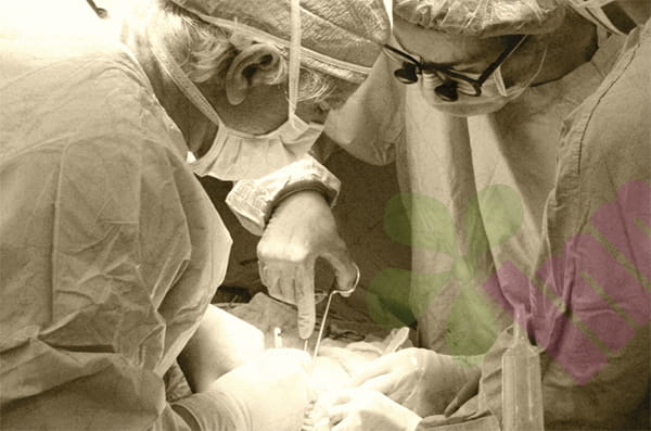






Several days to two weeks after surgery, the incision dressing becomes soaked with a pale yellow liquid. Upon removal, oily exudate can be seen at the incision edges. When the incision is squeezed, the liquid overflows from the suture gaps, but the surrounding skin shows no obvious redness or swelling, and the patient may not develop a fever. Clinically, this condition is called postoperative wound fat liquefaction. It is not a typical purulent infection, but rather a fluid accumulation formed when the subcutaneous fat layer undergoes ischemic necrosis after surgical trauma, causing fat cells to rupture, releasing lipids, and accumulating within the incision. Fat liquefaction itself is not a bacterial infection, but improper treatment can lead to secondary infection and prolong healing time.
How exactly does fat liquefaction occur?
When the skin and subcutaneous tissue are surgically cut, blood vessels within the fat layer are severed, reducing local blood supply. The high temperatures generated by electrocautery and electrocoagulation cause fat cells to rupture, releasing liquefied lipids. Obese patients have a thick subcutaneous fat layer and low vascular density per unit volume, making the fat tissue at the incision edge more prone to ischemia. After fat cell necrosis and liquefaction, fluid accumulates within the incision, forming cavities, and exudate flows out along the suture gaps or incision edges. The liquefied area lacks sufficient blood supply, making it difficult for white blood cells and antibiotics to reach it; therefore, simply administering medication or injections is unlikely to eliminate the liquefied cavities.
Does oozing yellow fluid mean there's an infection?
Both fat liquefaction and infection can present as incision effusion, but they are distinct. Fat liquefaction effusion is typically pale yellow, clear, or oily, with no noticeable odor. The surrounding skin is mildly red and swollen, and pain is not severe; the patient's temperature is usually normal. Infected effusion is often cloudy and purulent, yellowish-green, or bloody, with a foul odor. The area around the incision is markedly red, swollen, hot, and painful, and may be accompanied by fever and elevated white blood cell count. Culture of the effusion can aid in diagnosis. Fat liquefaction cultures often show sterile growth or only a small number of bacteria, while infected cultures will cultivate pathogenic bacteria. Do not mistake all fat liquefaction for infection, nor should infectious effusion be mistaken for ordinary liquefaction.
Who is more prone to fat liquefaction?
Patients with obesity, thick subcutaneous fat layers, poorly controlled diabetes, hypoalbuminemia, anemia, long-term use of glucocorticoids, malnutrition, prolonged surgery time, frequent use of electrocautery, high incision tension, and frequent postoperative coughing have a higher risk of fat liquefaction. In diabetic patients, microvascular complications further impair blood supply to the fat layer, and high blood sugar also suppresses leukocyte function, making liquefaction highly susceptible to infection. In obese patients, the large dead space within the fat layer after incision closure hinders exudate drainage, also contributing to persistent liquefaction.
Why is the incision opened when changing dressings?
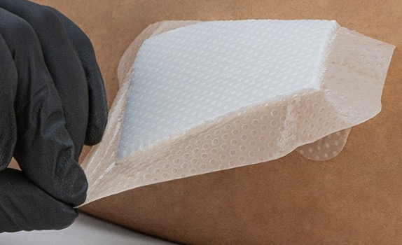
After fat liquefaction, the fluid accumulates under the skin. If only the outer dressing is changed, the fluid cannot drain and will form a high-pressure cavity within the incision, spreading to surrounding tissues and potentially breaking through the deep fascia or leaking from other suture gaps. Doctors often remove some sutures to open the incision, allowing the liquefied fat and exudate to drain fully. This procedure may appear to enlarge the wound, but it's actually to establish proper drainage. After opening, the wound is rinsed with saline solution to remove necrotic fat fragments, and a drainage strip or negative pressure drainage is placed. An absorbent dressing is then applied. Dressings are changed daily when exudate is high, gradually increasing the interval as it decreases. Granulation tissue grows upwards from the base. Secondary suturing or allowing the wound to heal spontaneously is considered once the wound is clean and exudate is minimal.
What precautions should be taken when caring for a patient at home?
Do not squeeze the incision to try to expel fluid, as this may push the infection deeper. Do not repeatedly irritate the incision with alcohol, iodine, or hydrogen peroxide, as these disinfectants can irritate fatty tissue and newly formed granulation tissue. Do not heat the incision with a heat lamp, as this will dry the surface and close the drainage. Do not apply toothpaste, powders, or folk remedies. Keep the dressing dry and change it promptly if it becomes soaked with exudate. Observe the extent of redness and swelling around the incision, as well as changes in body temperature and pain. Patients with diabetes should monitor their blood sugar, ensure adequate protein intake, and supplement with vitamin C and zinc if necessary. If the exudate suddenly increases, develops an odor, the redness and swelling expands, or fever develops, seek medical attention immediately.
How to prevent and reduce fat liquefaction?
Preoperatively, control blood sugar and weight, and improve nutritional status. During surgery, avoid excessively high electrocautery power, and perform sharp dissection of the fat layer to minimize thermal damage. Suture in layers to eliminate dead space, and place subcutaneous drainage if necessary. Postoperatively, use an abdominal binder to reduce tension and avoid vigorous coughing and straining during bowel movements. Obese patients should engage in moderate activity in the early postoperative period to avoid sudden increases in incision tension. For patients with liquefaction, the key is adequate drainage, infection control, nutritional support, and patient dressing changes. Fat liquefaction is not an infection, but it requires wound treatment; it cannot be treated solely with antibiotics, nor can it be left to heal on its own. Most patients heal gradually within two to four weeks after drainage and dressing changes; a small number of patients with concurrent infection or deep fascial involvement require longer treatment and surgical intervention. For more information on Innomed® Silicone Foam Dressing, refer to the Previous Articles. If you have customized needs, you are welcome to contact us; You Wholeheartedly. At longterm medical, we transform this data by Innovating and Developing Products that Make Life easier for those who need loving care.
Editor: kiki Jia


