 English
 عربى
عربى Español
 русский
 中文简体
中文简体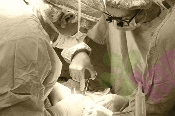


What exactly is Fournier gangrene?
Fournier gangrene is essentially a necrotizing fasciitis caused by a mixed bacterial infection. It is usually not caused by a single bacterium, but rather by the combined action of multiple bacteria, including Escherichia coli, Streptococcus, Staphylococcus aureus, and various anaerobic bacteria. These bacteria enter the subcutaneous tissue through small fissures in the perianal skin, ruptured hemorrhoids, infected periurethral glands, or local surgical incisions, and then spread rapidly along the fascial plane. The fascia is connective tissue that surrounds muscles and has a poor blood supply, allowing bacteria to multiply unimpeded by the immune system, while simultaneously producing various toxins and gases. Toxins damage local blood vessels, leading to tissue ischemia and necrosis; gases diffuse subcutaneously, forming subcutaneous emphysema. The entire infection spreads at an alarming rate, reaching one to two centimeters per hour. Literature reports that it can take as little as six to twelve hours from the appearance of skin erythema to the development of extensive tissue necrosis. Without timely surgical incision, drainage, and debridement, the infection can further lead to sepsis, septic shock, and multiple organ failure.
Who are most susceptible to Fournier gangrene?
Fournier gangrene is extremely rare in healthy individuals, and the vast majority of patients have underlying medical conditions. Diabetes is the most significant risk factor, with over 60% of patients with Fournier gangrene having a history of diabetes. Prolonged hyperglycemia impairs white blood cell function, reducing the body's ability to fight infection; simultaneously, the hyperglycemic environment itself provides an excellent culture medium for bacteria. Patients who are bedridden for extended periods or suffer from urinary or fecal incontinence are also at high risk, as the perineum is constantly exposed to a moist environment contaminated with excrement, leading to a weakened skin barrier function and increased susceptibility to bacterial invasion. Obese patients have thicker subcutaneous fat and relatively poorer tissue blood supply, making them more susceptible to the spread of infection once it occurs.
What are the early signs of Fournier gangrene?
Early identification of Fournier gangrene is crucial for saving lives. A typical early symptom is sudden pain in the perineum or perianal area. This pain differs from that of ordinary hemorrhoids or folliculitis; it is a deep, persistent, throbbing pain. Patients often cannot pinpoint the exact location of the pain, only feeling discomfort throughout the perineal area. Within hours of the onset of pain, the local skin begins to redden and swell, with indistinct borders, unlike cellulitis which has a clear red line. As subcutaneous gas develops, gently pressing the red and swollen area with a finger will produce a peculiar squeezing sensation, like squeezing a sponge containing tiny air bubbles. This is a typical sign of subcutaneous emphysema and an important diagnostic criterion for Fournier gangrene. Patients will also experience systemic poisoning symptoms, including high fever reaching 39 or even 40 degrees Celsius, chills, rapid heartbeat, shortness of breath, lethargy, and complete loss of appetite. If medical attention is not sought at this stage, after several to over ten hours, the red and swollen skin will turn purplish-brown, then blisters, necrosis, and ulceration will occur, releasing a foul-smelling, watery exudate. This process is very rapid; from the initial symptoms to skin necrosis, most cases are completed within 24 to 48 hours.
How is Fournier gangrene treated?
Treatment of Fournier gangrene requires swift action, with surgical debridement being the core procedure. Once diagnosed, patients are typically taken to the operating room within hours. Doctors will incise along all boundaries of the infected area, thoroughly removing necrotic fascia, fat, and skin until fresh, bleeding healthy tissue is reached. This surgery can result in significant tissue loss; large areas of the perineum, scrotum, and perianal skin may be removed, sometimes even requiring the removal of part of the testis or anal sphincter. The wound remains open after debridement and cannot be sutured, as suturing would trap bacteria, allowing them to multiply. Post-operatively, the wound needs to be rinsed two to three times daily with copious amounts of saline or diluted povidone-iodine, while systemic broad-spectrum antibiotics are administered to cover both aerobic and anaerobic bacteria. Debridement is often not a one-time procedure; patients need to be re-evaluated 24 to 48 hours post-surgery to check for new necrotic tissue and any missed areas. This may require two to three or even more debridement sessions until the wound base is completely clean and free of necrotic tissue.
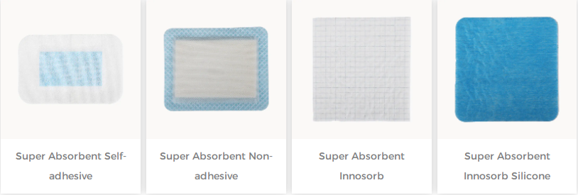
For more information on Innomed®Silicone Foam Dressing, refer to the Previous Articles. If you have customized needs, you are welcome to contact us; You Wholeheartedly. At long-term medical, we transform this data by innovating and developing products that make life easier for those who need loving care.
Editor: kiki Jia


