
 English
 عربى
عربى Español
 русский
 中文简体
中文简体






Patients with chronic wounds and diabetic foot ulcers often find that the skin around the wound edges becomes white and thick during dressing changes, forming a raised ring of keratin. In some cases, the keratin at the edges even rolls inward, covering the wound edge. This condition is professionally known as hyperkeratosis of the wound margin or calloused margin, and its formation is related to long-term immersion in exudate around the wound, friction from dressings, and excessive proliferation of epidermal cells during the repair process. Faced with this ring of keratin, many people's first reaction is to trim it off with scissors, thinking that smoothing it out will help the wound heal better. However, improper timing and method of trimming will not only fail to help healing but may also create new skin damage and entry points for infection. Therefore, whether to trim, who should trim, and how to trim need to be determined based on the wound type and the condition of the keratin.
How does the buildup of keratin at the edges of a wound form?
The stratum corneum of normal skin is in a dynamic balance of continuous generation and shedding, with the outermost layer of keratinocytes periodically shedding and being replaced by new cells. During wound healing, the proliferative activity of epidermal cells at the wound edge significantly increases, and keratinocytes rapidly divide and migrate, attempting to cover the wound. If this proliferative activity is faster than the normal shedding rate of the stratum corneum, it will accumulate at the wound edge, forming visible thickening. Simultaneously, wound exudate continuously soaks the edge skin, causing the stratum corneum to absorb water and swell, loosening the connection between it and the underlying living cell layer. Shed keratinocytes cannot leave the skin surface in time but instead adhere and accumulate in their original positions. Repeated friction and removal of dressings at the edges also exert mechanical stimulation on the skin around the wound; the more frequent the stimulation, the stronger the epidermal proliferative response. Thickening of the keratin at the edges of foot ulcers is particularly prominent in diabetic patients. This is related to the abnormal keratinization of the skin itself in diabetic patients and the repeated pressure on the soles of the feet. Ulcers are often hidden deep beneath thick calluses, and without removing some of the keratin, the true boundary of the ulcer may not even be visible.
In what situations does this layer of keratin need to be treated?
Excessive keratinization at the wound edge, if left untreated, can cause several practical problems. The thickened keratin ring acts like a dam above the wound surface, hindering the migration of epithelial cells from the edge to the center. Cells encounter physical obstacles and stall at the edge of the keratin layer, delaying wound closure. Beneath the keratin ring lies a moist groove where exudate and bacteria can easily accumulate, creating hidden corners prone to recurrent infection. The thick keratin also interferes with wound measurements, blurring the visible boundaries and causing discrepancies between the measured area and the actual defect. Therefore, trimming is necessary in the following situations: the keratin ring is significantly higher than the skin plane and rolls inward to cover the wound edge; fluid or purulent discharge is visible beneath the keratin; the thickness of the keratin affects the adhesion of the dressing to the wound edge, leading to leakage; and large calluses covering the ulcer opening around diabetic foot ulcers. If the stratum corneum only shows slight thickening, with smooth edges and no curling or covering, and wound measurement and dressing application are not affected, it can be left untreated for the time being. The focus should be on controlling exudation and reducing friction and irritation.
Why can't you trim your skin at home by yourself?
Trimming the edges of a wound is fundamentally different from routinely removing calluses from the hands and feet. Beneath the skin at the wound edge lies fragile tissue undergoing repair. The boundary between the stratum corneum and living tissue becomes unclear after exudation, making it difficult for non-professionals to determine which layer is safe to cut. Poor control of the force and direction of the scissors can easily cut through the epidermis and dermis beneath the stratum corneum while trimming the keratin, creating new areas of damage. These new damages are often next to the original wound, and the two wounds may merge and enlarge. Household scissors are not sterilized, and bacteria on their surface can inoculate the damaged areas at the wound edges, significantly increasing the risk of infection. Patients with diabetic foot ulcers have reduced sensation in their feet; they may not feel pain even if they cut too far and injure normal tissue. Combined with insufficient blood supply and immune defense in the lower limbs, a single improper trimming can lead to a serious spread of foot infection. Therefore, the removal of keratin from the wound edges should be performed by wound care professionals or trained physicians using sterile instruments and debridement techniques, carefully cutting thin layers of keratin while clearly seeing the boundary between the keratin and living tissue.
What are some methods for exfoliating?
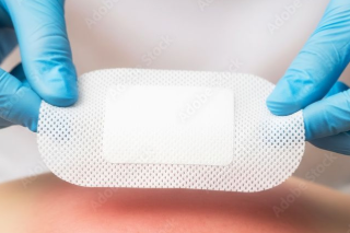
Clinically, methods for treating keratinized wound edges include three categories: sharp instrument debridement, mechanical exfoliation, and autolytic debridement. Sharp instrument debridement uses a sterile scalpel to remove a thin layer of keratin along the direction of keratin proliferation, stopping when the keratin is close to normal skin thickness, leaving a very thin layer of keratin to avoid damaging the granular layer. After treatment, the edges are cleaned with saline solution, and any bleeding is observed. If pinpoint bleeding occurs, it indicates that the keratin has been removed to the papillary dermis, requiring immediate pressure and wound protection. Mechanical exfoliation is suitable for cases of thick but well-defined keratin. Sterile forceps are used to gently open the keratinized edge, and sterile cotton swabs are used to peel off the loosened keratin scales. Adhesive areas should not be forcibly pulled. Autolytic debridement is suitable for mild keratin thickening or cases where the patient cannot tolerate sharp instruments. Hydrogel is applied to the keratinized ring, and then sealed with a dressing. Hydration softens the keratin, allowing it to break off naturally. This process is slower but less invasive. After treatment, a skin protectant may need to be applied to the wound edges to reduce re-keratinization. In subsequent dressing changes, a soft silicone dressing with a large edge adhesion area should be used to reduce friction and irritation that could induce hyperkeratosis. The principles for treating keratinized wound edges can be summarized as follows: anything affecting healing must be treated; treatment must be performed by a professional; and the method should be chosen to minimize damage and avoid creating new wounds. This order cannot be reversed. For more information on Innomed® Silicone Foam Dressing, refer to the Previous Articles. If you have customized needs, you are welcome to contact us; You Wholeheartedly. At long-term medical, we transform this data by innovating and developing products that make life easier for those who need loving care.
Editor: kiki Jia


