 English
 عربى
عربى Español
 русский
 中文简体
中文简体


When changing dressings, opening the wound reveals red, moist granulation tissue resembling small strawberries – a good sign of healing. However, if the granulation tissue becomes excessively bright red or dark red, is watery, feels soft to the touch, or even protrudes above the surrounding skin, caution is advised; it may be granulation edema. Adding to the problem, granulation edema and early infection can sometimes look very similar, but their treatment approaches are completely different. Treating edema as an infection with anti-inflammatory drugs, or treating an infection as edema with dehydration, will both lead to misguided wound healing.
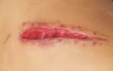
What does granulation tissue edema look like?
Normal granulation tissue should be bright red or pink, moist but not excessively so, with uniform granules. A light touch with a finger will cause a few pinpoint bleeding, indicating viability. When granulation tissue is edematous, the color becomes excessively bright red or dark red, the surface becomes shiny as if soaked in water, and the granules become flat or merge into a single mass. The most typical manifestation is that the granulation tissue protrudes above the wound base, even bulging from the wound edge, feeling soft and elastic, like a swelled sponge. Pressing on it will leave a small indentation, which slowly recovers upon release. This is due to excessive fluid accumulation between tissues, causing an imbalance in extracellular fluid osmotic pressure. While edematous granulation tissue grows in abundance, its quality is poor; it is brittle, prone to bleeding, and hinders the surrounding epithelial cells from moving towards the center, thus delaying wound closure.
What does an infection look like?
Granulation tissue in infected wounds differs from edema. In the early stages of infection, granulation tissue may remain red, but its color is darker or purplish, covered with a thin, grayish-white or yellowish-white membrane. As the infection worsens, the granulation tissue becomes fragile, bleeding or crumbling at the slightest touch. Wound exudate increases and changes in its characteristics, changing from a clear pale yellow to a cloudy yellowish-white or yellowish-green. Another typical sign of infection is odor; the decomposition of necrotic tissue by bacteria produces a putrid or ammonia-like smell. The surrounding skin will become red, swollen, warm, and tender; in severe cases, the redness and swelling may extend beyond the area covered by the dressing. Patients may experience low-grade fever or general malaise, but this may be less noticeable in the elderly or those with weakened immune systems.
How to identify it? Here are a few key points :
First, observe the color and texture of the granulation tissue. Edema-related granulation tissue is bright red or dark red, plump, shiny, and raised above the wound surface, but its texture is soft and elastic, neither brittle nor fragile. Infected granulation tissue is dark purple or grayish-white, with a membranous covering on the surface, and its texture is fragile; it bleeds or falls off easily with a gentle scrape with a cotton swab. Second, observe the exudate. Edema-related exudate is copious but clear, mostly a pale yellow, serum-like fluid, without a noticeable odor. Infected exudate is cloudy, thick, yellowish-green, or streaked with blood, and has a distinct fishy or putrid odor. Third, observe the surrounding skin. Edema-related wound edges usually do not show obvious inflammatory reactions; they may simply be whitish from being soaked in exudate. Infected wound edges will definitely be red, hot, and swollen, with a clear inflammatory boundary. Fourth, observe the systemic reaction. Simple edema rarely causes fever or elevated blood cell counts, while infection, as it progresses, will cause a systemic inflammatory response.
A simple functional test can also be performed. Gently press the surface of the granulation tissue with a sterile cotton swab. Edema-filled granulation tissue will indent when pressed and slowly spring back when released; infected granulation tissue may rupture and bleed directly when pressed, or the pus underneath may be squeezed out. For deep infections, a sterile probe or fine cotton swab can be used to gently probe the base of the wound. If a deep cavity is detected or a hard bone surface can be felt, be alert to osteomyelitis or a deep abscess.
The handling methods are completely different.
The key to managing granulation tissue edema is reducing interstitial edema. First, check the wound for undermining cavities or sinuses that might obstruct drainage. If present, expand the drainage area or place a drainage tube. Locally, apply hypertonic saline gauze as a wet dressing; the hypertonic environment draws fluid from the interstitial spaces. Specifically, soak sterile gauze in 3-5% hypertonic saline, wring it until it's no longer dripping, and gently apply it to the edematous granulation tissue. Cover with dry gauze and change the dressing once or twice daily. The edema should significantly decrease after two to three days. If the edema recurs, consider systemic factors such as hypoproteinemia, heart failure, or renal insufficiency; blood tests are necessary for these conditions. The key to managing infection is anti-infection treatment and debridement. Superficial infections can be treated with silver ion dressings or povidone-iodine gauze as wet dressings, changing the dressing daily. If the infection has penetrated the fascia layer or formed an abscess, incision and drainage or surgical debridement are required; this cannot be done at home. For more information on Innomed® Silicone Foam Dressing, refer to the Previous Articles. If you have customized needs, you are welcome to contact us; You Wholeheartedly. At long-term medical, we transform this data by innovating and developing products that make life easier for those who need loving care.
Editor: kiki Jia


