 English
 عربى
عربى Español
 русский
 中文简体
中文简体


When changing the wound dressing, the wound surface looks relatively clean, with no obvious pus, and the surrounding area is neither red nor swollen, but it just won't heal. Even after weeks or months of dressing changes, the exudate remains the same, granulation tissue just won't grow, and the epithelium can't spread. This situation is very likely caused by a biofilm. A biofilm is an extracellular polymer secreted by bacteria after adhering to the wound surface, forming a mucous-like protective layer that encapsulates themselves. It differs from free-floating bacteria, which are easily cleared by antibiotics and immune cells; bacteria within a biofilm are encapsulated by extracellular polymers, like wearing a bulletproof vest, preventing antibiotics from penetrating and immune cells from engulfing them. Adding to the problem, biofilms are difficult to see directly with the naked eye; they may only be a thin, translucent membrane adhering to the wound surface, blending in with the normal, moist wound base, and easily overlooked.
What are the criteria for identifying biofilms?
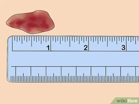
Although invisible, biofilms have several key indicators. The first is stagnation of healing. A wound, after standard dressing changes, shows no obvious signs of infection, necrotic tissue residue, or systemic malnutrition, but if it shows no signs of shrinking for two to four weeks, this stagnation strongly suggests a biofilm. The second is altered exudate characteristics. With a biofilm, the exudate is often more viscous and has increased stringiness, unlike the thin, normal serum-like exudate. Sometimes the exudate is pale yellow or pale green and gelatinous, and can be pulled into thin threads when swabbed with a cotton swab. The third is a mild, persistent inflammatory response. Biofilms do not cause severe redness, swelling, heat, or pain, but there is often a faint red halo around the wound, and the skin temperature is slightly higher than the opposite side. This low-grade inflammation is poorly treated with antibiotics, and recurrence occurs upon discontinuation. The fourth is poor response to antibacterial treatment. After using topical antibacterial dressings or systemic antibiotics, the wound may temporarily improve, but it quickly returns to its original state after discontinuation. This is because the medication kills the surface bacteria, but the deep biofilm structure remains intact.
Another commonly used clinical method for auxiliary assessment is the swab rolling test. Gently roll a sterile cotton swab over the wound surface, rather than rubbing vigorously. If there is a mature biofilm on the wound, the swab will pick up a layer of transparent or pale yellow mucus film after rolling, adhering to the swab like glue. Ordinary exudate is watery and will not form a film on the swab. This method is simple to perform, requires no special equipment, and can be done in outpatient settings or at home.
How can we confirm the presence of biofilms?
If a biofilm is highly suspected clinically, it can be confirmed through pathogen testing. The most common method is wound secretion culture combined with biofilm detection. Routine bacterial culture only indicates the presence of bacteria, not whether they exist as a biofilm. Specialized biofilm-related tests, such as fluorescent staining or electron microscopy, are required. Another approach is molecular biology testing, such as polymerase chain reaction (PCR) or next-generation sequencing (NGS), which can directly detect biofilm-related genes, such as those responsible for synthesizing extracellular polysaccharides. These tests are relatively expensive and are generally used for complex cases or research. In routine clinical practice, clinical diagnoses are more often based on healing stagnation and response to conventional treatments, rather than waiting for test results.
What to do after discovering a biofilm?
Once a biofilm is confirmed or highly suspected, conventional dressing changes are insufficient. Managing biofilms requires a multi-pronged approach. The core of this approach is physically disrupting the biofilm structure. The extracellular polymers of biofilms are mainly polysaccharides, proteins, and extracellular DNA, which can be broken down by specific enzymes. Clinically, alginate dressings are commonly used; the calcium ions they release can chelate calcium ions in the biofilm, disrupting its stability. Dressings containing EDTA are also used; EDTA can chelate divalent cations, disrupting the structural integrity of the biofilm. A more direct method is debridement. Using a scalpel, curette, or pulse irrigation device, the biofilm attached to the wound surface, along with the underlying thin layer of tissue, is removed. After debridement, fresh tissue is exposed at the base of the wound, and the biofilm is completely removed. It is important to note that a single debridement may not remove all biofilm, as it may penetrate deep into the interstitial spaces, requiring multiple debridements. After each debridement, an anti-biofilm dressing, such as one containing silver ions or iodine, should be applied immediately to prevent biofilm reformation within 24 hours. The frequency of dressing changes should also be increased, as biofilm formation takes time, generally within 48 to 72 hours to form a mature biofilm structure. If the dressing is changed before the biofilm matures, bacteria do not have enough time to establish a protective layer and are easily removed. For wounds diagnosed with biofilm infection, it is recommended to change the dressing daily, or even twice a day, to prevent the biofilm from reforming.
Is prevention more important than treatment?
Once a biofilm forms, it becomes very difficult to manage, so prevention is more important than treatment. The core of prevention is interrupting the early stages of biofilm formation. The first six hours after bacteria attach to the wound are a critical window. During this stage, bacteria have not yet secreted large amounts of extracellular polymers, their structure is loose, and they are easily washed away or killed by antibacterial dressings. Therefore, in patients at high risk of biofilm formation, such as those with chronic wounds like diabetic foot ulcers, pressure ulcers, and venous ulcers, dressing changes must be frequent enough. Wounds with heavy exudate should be dressed daily, and wounds with less exudate should be dressed at least every other day. Each dressing change should involve rinsing the wound with a sufficient amount of saline solution to wash away airborne bacteria. The rinsing pressure should not be too low; at least four to seven pounds per square centimeter are needed to effectively remove loosely attached bacteria. This pressure can be generated using a 20 ml syringe with an 18-gauge needle. After rinsing, apply an anti-biofilm dressing. In addition, controlling wound exudate is also crucial, as exudate is a nutrient source for bacteria; the more exudate, the faster the biofilm grows. Using highly absorbent foam dressings or alginate dressings to promptly absorb exudate and keep the wound moderately moist but not waterlogged can significantly delay biofilm formation. For chronic wounds with recurrent biofilm infections, silver ion-containing foam dressings can be considered for daily maintenance therapy. Silver ions can continuously inhibit bacterial activity, and even if a biofilm forms, its structure is less dense than ordinary biofilms, making it easier to remove. For more information on Innomed® Silicone Foam Dressing, refer to the Previous Articles. If you have customized needs, you are welcome to contact us; You Wholeheartedly. At long-term medical, we transform this data by innovating and developing products that make life easier for those who need loving care.
Editor: kiki Jia


