 English
 عربى
عربى Español
 русский
 中文简体
中文简体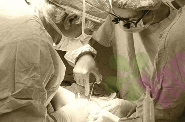


In later stages, pressure ulcers, especially in bony prominences like the sacrum, coccyx, or heels, sometimes form a hard, black scab that is dry and hard, resembling armor. Many people see this scab and think it's a protective layer to prevent bacteria from entering, so they want to leave it. Others believe it's necrotic tissue and should be removed. This issue is indeed controversial in clinical practice, and the key is to consider the type of scab and the patient's overall condition.
There are two types of black scabs :
The black scabs on pressure sores are not the same thing; they can be divided into dry gangrene and wet gangrene. Dry gangrene is caused by insufficient arterial blood supply, leading to tissue ischemia and necrosis. The necrotic tissue dries and hardens after the water evaporates, turning black in color, with clear borders, no obvious redness or exudation around it, and no odor. This black scab may act as a protective layer for the patient, sealing the underlying tissue and preventing external bacteria from entering. However, if the patient has very poor arterial blood flow in the lower limbs, with cold, purplish feet and no palpable pulse, cutting off the black scab will only expose a dry, ischemic, and difficult-to-heal wound, and may even lead to infection.
Wet gangrene is different. Beneath this black scab lies necrotic tissue, pus, and a large number of bacteria. The scab isn't hard to the touch; it's soft and flaky, and you might feel a fluid fluctuation when you press it. Yellow or green exudate often oozes from the edges, emitting a foul odor. The surrounding skin is red, swollen, and warm; the patient may also have a fever. This type of scab must be cut; otherwise, the pus won't drain, and bacteria will spread along the fascia, leading to sepsis and even death. Most pressure sores requiring treatment in clinical practice fall into this category.
What needs to be assessed before cutting?
Before deciding whether to remove the scab, several crucial assessments cannot be skipped. First is a blood supply assessment. Feel for the pulse of the dorsalis pedis and posterior tibial arteries, then press the skin next to the scab with your fingers and observe the speed of color recovery. Normally, the skin turns white after pressing and turns red within one to two seconds after releasing the pressure. If it takes more than five seconds to return to normal, it indicates severe arterial blood supply insufficiency. If possible, an ankle-brachial index (ABI) or lower limb vascular ultrasound will provide a more accurate result. If the ABI is less than 0.5, it indicates severe ischemia, and handling the scab in this case requires extreme caution. Second is assessing for infection beneath the scab. The simplest method is to gently pry up a small corner of the scab with a sterile needle to see if any fluid drains. If the fluid is clear and serum-like, it's generally not a problem; if the fluid is cloudy, yellow, or bloody pus with a foul odor, it indicates infection and requires incision and drainage. Third is assessing the patient's overall condition. A patient with severe heart failure, kidney failure, or advanced cancer may not be able to withstand a thorough debridement surgery. In such cases, it is better to be conservative and only perform surface debridement and drainage, rather than extensive resection.
How do I cut off the black scab that needs to be removed?
Once you've identified the eschar that needs to be removed, never try to cut it yourself at home with scissors; this is absolutely forbidden. The tissue layers beneath a pressure ulcer are unclear; it may have already rotted into the muscle or even bone. Cutting it yourself can easily damage deep, healthy tissue or leave incomplete removal, leaving dead zones. The correct procedure is to do it in a hospital operating room or debridement room. The doctor will soak the eschar in iodine or saline solution for five to ten minutes to soften it slightly. Then, using sterile forceps and tissue scissors, they will remove the necrotic tissue layer by layer along the boundary between the eschar and normal tissue. During the process, the viability of the tissue must be constantly assessed, and the procedure should continue until fresh bleeding tissue is reached. Sometimes, what appears to be a small eschar may reveal a deep necrotic cavity upon incision, requiring the removal of a much larger area than initially thought. After removal, the wound remains open and cannot be sutured, as residual bacteria and exudate must continue to drain. The wound will then be rinsed with saline solution, and an appropriate dressing will be chosen based on the amount of exudate, such as alginate dressings or silver foam dressings, before covering it with dry gauze. After the surgery, the dressing needs to be changed once or twice a day to observe whether new necrotic tissue appears on the wound. If so, it needs to be cleaned again.
How to care for black scabs that shouldn't have been cut?
For dry gangrene-type eschars, especially ischemic pressure ulcers, the treatment principle is completely opposite. Do not cut, do not soak, and do not use any ointments that attempt to soften or dissolve the eschar. The goal is to keep the eschar dry and intact to prevent it from becoming wet gangrene. Gently wipe the surface of the eschar once a day with alcohol or povidone-iodine to disinfect and sterilize. Do not use oily ointments or hydrogel dressings, as these will soften and moisten the eschar, making it easier for bacteria to grow. Protect the normal skin around the eschar by applying zinc oxide ointment or a skin protectant cream to prevent maceration from urine and feces. The patient should be turned frequently, changing position every two hours to avoid continued pressure on the eschar area. At the same time, actively improve the patient's overall condition, treat the primary disease, correct anemia and hypoproteinemia, and consult a vascular surgeon if necessary to assess whether interventional or bypass surgery is possible to improve blood supply. If the eschar remains dry, odorless, and without surrounding redness and swelling under conservative care, it can exist for a long time without causing problems. However, once the scab softens, becomes wet, starts to ooze fluid, or emits a foul odor, it indicates that it has changed from dry to wet. At this point, it is too late to be conservative and immediate incision and drainage are necessary.
A special exception
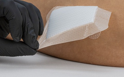
Stable, dry eschar on the heel deserves special mention. Some studies suggest that if a patient is unable to walk, the eschar on the heel is dry, firm, without surrounding redness or oozing, and ultrasound examination reveals no underlying fluid accumulation or abscess, this eschar can be preserved and does not necessarily need to be removed. This is because the blood supply to the heel is inherently poor, and the wound exposed after removing the eschar is difficult to heal and may even remain open for a long time. In comparison, retaining this natural protective layer is safer. However, regular follow-up observation is necessary, with a doctor's evaluation approximately every two weeks. Be prepared to cut it open immediately if any signs of infection appear. In general, the key to determining whether or not to remove pressure eschar is not the eschar itself, but the condition underneath. Dry, stable, and uninfected eschars can be preserved for protection; wet, fluctuating, and infected eschars must be incised and drained. This judgment is best made by an experienced wound therapist or doctor; family members should not make decisions at home based on their intuition. For more information on Innomed® Silicone Foam Dressing, please refer to the Previous Articles. If you have customized needs, you are welcome to contact us; you wholeheartedly. At long-term medical, we transform this data by innovating and developing products that make life easier for those who need loving care.
Editor: kiki Jia


