 English
 عربى
عربى Español
 русский
 中文简体
中文简体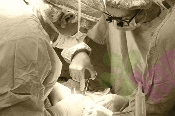


Skin grafting involves taking a healthy piece of skin from the patient and transplanting it to a burn, traumatic, or surgically unhealed wound. This harvested skin is called a skin graft, and unlike a skin flap, it doesn't have its own blood vessels. After being transplanted to its new location, the skin graft itself has no blood supply; it relies on tissue fluid seeping from the wound base for nutrition, while waiting for capillaries from the wound to slowly grow into the graft. This process usually takes five to seven days. During this time, any slight relative movement between the skin graft and the wound base can cause the newly growing capillary buds to break, and hematomas or seromas beneath the graft can also push it up, preventing it from contacting tissue fluid. Once the skin graft detaches from the base, it will become ischemic and necrotic, and the entire skin graft will fail. Therefore, the most important post-operative care principle for skin grafts is immobilization, keeping the graft area firmly fixed and completely still.
How did the leather pieces survive?
To understand the importance of immobilization, it's essential to first understand the skin graft survival process. On the first two days after surgery, the skin graft floats on a substrate, supported by blood-like fluid seeping from the wound. At this stage, the graft is merely lightly attached to the wound without any firm connection. From the third to the fifth day post-surgery, capillaries begin to grow into the skin graft; this stage is called vascular recanalization. These capillary buds are extremely delicate, each only a few micrometers in diameter. If the skin graft slips, these capillary buds will be torn off at the root.
Which parts require the most stringent braking?
The immobilization requirements differ depending on the location of the skin graft. Skin grafts to the limbs are relatively easier to control because joints can be immobilized with casts or splints. For example, after a skin graft to the lower leg, a long leg cast from the knee to the ankle is needed to immobilize both the ankle and knee joints, preventing flexion and extension of the ankle and thus preventing the skin graft from being pulled by skin movement. Skin grafts to the hand require palmar or dorsal plaster casts to immobilize the fingers in a functional position, preventing the skin graft from folding or stretching during finger movement. Immobilization of skin grafts to the head, face, and torso is more difficult because people constantly breathe, eat, and turn their heads—these everyday actions all pull on the surrounding skin.
How can effective immobilization be achieved ?
Immobilization is not just a theoretical concept; it requires specific tools and methods. The most common methods are plaster casts or polymer splints, applied on the first day after surgery to fix the limb containing the skin graft in a position where the skin graft is not under tension. For example, for a skin graft on the front of the thigh, the knee joint needs to be straightened and immobilized, not bent. For a skin graft on the back of the thigh, the knee joint needs to be flexed at about 30 degrees to allow the skin on the back to loosen. For a skin graft on the back of the hand, the fingers need to be straightened or slightly flexed, depending on the position of the skin graft. After plaster immobilization, it is necessary to regularly check whether the edges of the plaster are rubbing against the skin and whether the plaster itself is loose. If the plaster becomes loose, the skin graft can slide inside, negating the purpose of immobilization .
Besides braking, what else should we pay attention to?
Several issues need to be addressed simultaneously during immobilization. One is hematoma or seroma under the skin graft. Even with strict immobilization, fluid may still accumulate under the graft. If left untreated, this fluid can push the graft up, causing it to float and necrose. The solution is to make several small incisions in the graft or puncture the edge with a needle 24 to 48 hours post-surgery to drain the fluid; this is called fenestration drainage. The small holes in the graft will heal on their own within a few days and will not affect healing. Another issue is bleeding and infection at the graft edge. The junction between the graft and normal skin is the most prone to problems; carefully observe this edge for redness, swelling, pus, or graft lifting during dressing changes. If the graft edge becomes black, dry, and hardened, it indicates that a small portion of the graft has necrotized and needs to be trimmed away promptly to prevent it from becoming a culture medium for bacteria.
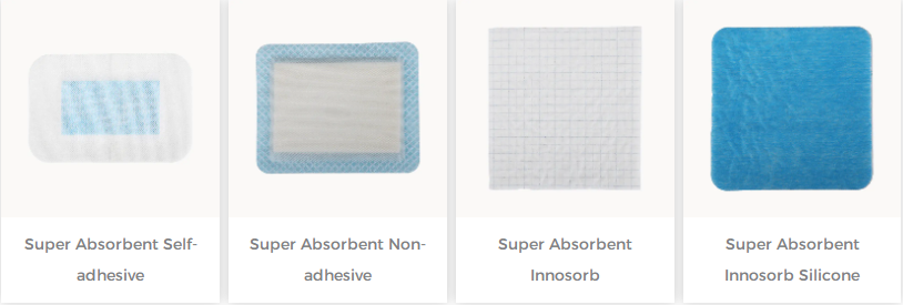
For more information on Innomed® Silicone Contact Layer, refer to the Previous Articles. If you have customized needs, you are welcome to contact us; You Wholeheartedly. At long-term medical, we transform this data by innovating and developing products that make life easier for those who need loving care.
Editor: kiki Jia


