 English
 عربى
عربى Español
 русский
 中文简体
中文简体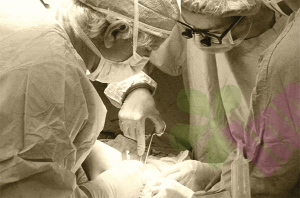


Wound healing is not a chaotic mass of cells randomly packed together, but a continuous process strictly ordered along a timeline. Different types of cells enter the wound area in sequence at different stages, each completing its specific task before exiting or transforming into the next form. This strict temporal arrangement resembles a fixed workflow. Understanding this sequence helps patients determine which stage their wound is currently in and clarifies why certain behaviors are prohibited at specific times.
Hemostasis phase :
The first step is the intervention of inflammatory cells, which occur within seconds to minutes after injury. Platelets are the first to respond; upon contact with exposed collagen fibers, they rapidly adhere, aggregate, and release their contents, forming a temporary embolus to block the ruptured blood vessel. Simultaneously, the coagulation cascade is activated, converting fibrinogen into fibrin. These fibrin molecules cross-link into a network structure, capturing platelets and red blood cells to form a blood clot. This clot not only stops bleeding but also serves as a temporary scaffold for subsequent cell migration. So, does wound repair begin immediately after hemostasis? Not at all; inflammatory cells are the second group to enter.
The inflammatory response peaks within hours to two days after injury. Neutrophils are the first white blood cells to migrate from the blood vessels into the wound tissue. Guided by chemokines, they aggregate towards the wound center, their main task being to engulf and digest bacteria and debris released from tissue damage. The number of neutrophils peaks 24 to 48 hours after injury, then gradually decreases through apoptosis. Monocytes enter and transform into macrophages within two to three days after injury. Macrophages not only continue to clear residual pathogens and necrotic material but also secrete various signaling molecules that recruit the next wave of cells and regulate their activity. If a significant increase in wound exudate is observed during this stage, and its color turns yellowish-green, it usually indicates that the bacterial load exceeds the clearance capacity of neutrophils, which is an abnormal signal.
Proliferation stage :
The process begins around the third day after injury and lasts for one to three weeks, during which fibroblasts become the primary working cells. Fibroblasts migrate from the wound edges and surrounding undamaged connective tissue towards the center, synthesizing and secreting large amounts of type III collagen and glycosaminoglycans to fill the volume of tissue defects. The newly synthesized collagen fibers initially exhibit a disordered arrangement and have significantly lower mechanical strength than normal skin. Simultaneously, basal cells at the wound edges begin to migrate towards the center; these epidermal cells need to cover the surface of newly formed granulation tissue to rebuild the epithelial barrier. In a moist environment, the migration rate of epidermal cells is significantly faster than in a dry environment because cell migration requires a sufficiently hydrated matrix. Clinicians emphasize keeping the dressing moist but not overly saturated during this stage precisely to create suitable conditions for the forward migration of epidermal cells.
Reshaping stage :
This is the last step in the process, starting three weeks after the injury and lasting for months or even over a year. During this long process, the number of fibroblasts decreases, and some transform into myofibroblasts, reducing the wound area through contraction. The previously deposited type III collagen is gradually replaced by type I collagen, and the arrangement of collagen fibers changes from a random state to an orderly arrangement along the tension lines. After reshaping, the tensile strength of the scar tissue can reach 70% to 80% of that of normal skin, but it cannot completely restore the original structure. So what can patients do at this stage? Keeping the wound site free from continuous traction and ultraviolet radiation are two effective interventions. Physical tension can stimulate excessive contraction of myofibroblasts, leading to scar hyperplasia, while ultraviolet radiation can disrupt the function of newly formed melanocytes, resulting in hyperpigmentation or hypopigmentation.
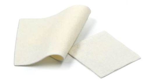
The sequence of these four stages is fixed; any stage being completed earlier or later will affect the overall progress. For example, in diabetic patients, macrophage function is impaired, prolonging the inflammatory phase and causing dysregulation in the concentration of signaling molecules, preventing fibroblasts from entering and functioning normally on time. This is one of the direct causes of delayed wound healing in diabetic patients. In a state of insufficient local blood supply, the supply of oxygen and nutrients is limited, reducing the collagen synthesis rate of fibroblasts, which also prolongs the time required for the proliferation phase. At the treatment level, debridement may reset part of the inflammatory phase, but this is to remove necrotic tissue to shorten the overall time. Observing the color and exudate characteristics of the wound bed during dressing changes is essentially determining whether current cellular activity is within the normal timeframe. If the duration of a certain stage significantly exceeds the expected window, it indicates the presence of interfering factors requiring medical intervention. For more information on Innomed® Alginate Dressing, refer to the Previous Articles. If you have customized needs, you are welcome to contact us; You Wholeheartedly. At long-term medical, we transform this data by innovating and developing products that make life easier for those who need loving care.
Editor: kiki Jia


