
 English
 عربى
عربى Español
 русский
 中文简体
中文简体






Urine flows continuously from the stoma, unlike the intermittent discharge of feces in an enterostomy. This continuously moist working environment determines that the core challenges of urostomy care are completely different from those of enterostomy. The difficulty of enterostomy lies in the chemical corrosiveness of excrement and the mechanical damage to the rubber surface of the stoma by solid components, while the difficulty of urostomy lies in the combination of three factors: continuous urine immersion, urine crystal formation, and fungal infection. Urine itself contains urea, uric acid, and various electrolytes. When it comes into contact with the skin, urea is decomposed by bacteria on the skin surface to produce ammonia. The alkaline environment of ammonia disrupts the skin's acidic barrier. At the same time, phosphates and urates in urine easily precipitate under alkaline conditions, forming tiny crystals that deposit on the skin around the stoma. The sharp edges of these crystals cause physical friction damage to the skin, and the damaged, moist skin provides ideal growth conditions for fungi such as Candida.
Why is more precise cutting required for the urostomy base plate?
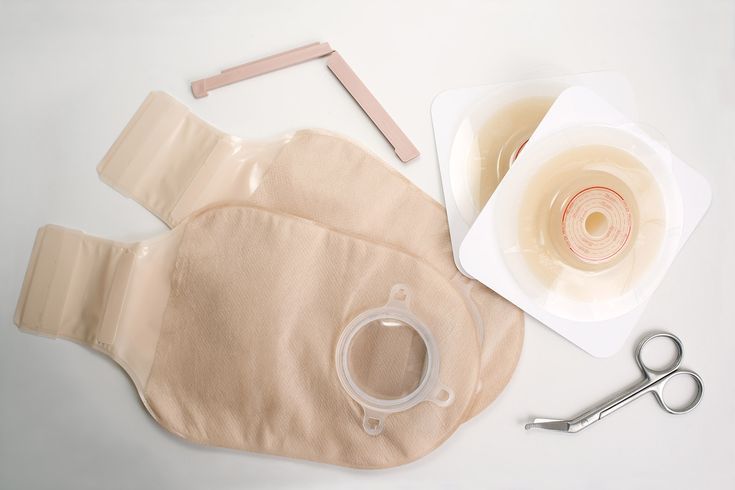
Urine from a urostomy is far more fluid than intestinal waste. Even a tiny gap of less than one millimeter between the baseplate and the stoma root can allow urine to seep beneath the baseplate within minutes. Since urine is colorless and transparent, unlike intestinal waste, there is no obvious color change indicating a malfunction. Often, a large accumulation of urine is only discovered when skin stinging and erythema appear. Therefore, the precision required for cutting a urostomy baseplate is much stricter than for an intestinal stoma. Ideally, the gap between the cut inner diameter and the stoma root should not exceed 0.5 millimeters. This gap is then sealed with a leak-proof ring. Stoma dimensions cannot be measured only once when the baseplate is replaced, as the stoma continues to shrink within six months post-surgery. Each baseplate replacement requires re-measuring the long and short diameters of the stoma root with a ruler, and the baseplate must be cut according to the actual measurements taken that month, not the old measurements from the previous month. The shape of the stoma may change at different stages after surgery, from the initial circle to an oval or irregular shape. When cutting, it is necessary to trim along the actual outline of the stoma to match the shape rather than mechanically cutting it into a perfect circle.
How can I ensure the adhesive surface is dry when replacing the chassis if urine continues to leak out?
This is the most skill-intensive part of urostomy care. The prerequisite for attaching the baseplate is that the skin in the attachment area must be absolutely dry. However, urine continues to leak from the urostomy during the change, and freshly dried skin can be soaked with newly expelled urine within seconds. Solving this problem requires attention to both pre-operative preparation and the rhythm of the procedure. Drinking as little water as possible one to two hours before the change can reduce urine output during the change, but complete water restriction is not necessary. Before starting the change, prepare all items, including warm-water-moistened gauze for cleaning, dry non-woven fabric, a leak-proof ring, a skin protectant, a pre-cut baseplate and ostomy bag, and a roll of clean gauze or a tampon. After cleaning the skin around the stoma, quickly pat it dry with gauze. Then, gently press a small roll of dry gauze or a tampon against the stoma opening to absorb the urine that continues to seep out during the change, providing a temporary two to three-minute window of relative dryness. The skin protective film is quickly sprayed and dried within this window. Once dry, any minor urine leakage is contained outside the film and does not affect the base's adhesion. After attaching the leak-proof ring and base, press firmly with your palm for three to five minutes. Body heat helps the adhesive soften and fill the skin texture, creating a strong seal. The goal for the entire process is to complete the application and pressing from removing the temporary absorbent to fully attaching the base within three minutes. This speed can be effectively achieved by pre-arranging all items in the order of use.
How to identify and treat common skin problems around a urostomy?
Several common abnormalities of the skin around a urostomy site each have their characteristic appearance. Candida infection presents as well-defined erythema in the area covered by the stoma, with scattered small red papules or even pustules. The edges are often surrounded by fine desquamation, accompanied by intense itching. The typical distribution area perfectly matches the stoma's adhesive surface. Irritant dermatitis caused by urine leakage differs in appearance from candida infection. The erythema has irregular, map-like borders, the skin surface is moist, and may even be eroded and oozing. Pain is severe, while itching is relatively mild. Urinary crystal deposits are visible to the naked eye as small white or grayish-white granules on the skin around the stoma. They can be wiped away with a damp gauze, and pinpoint bleeding may appear on the underlying skin after wiping. Pseudoverrucous hyperplasia is a benign proliferative change in the skin caused by long-term urine immersion. Grayish-white or skin-colored cauliflower-like papules appear around the stoma; they are firm, painless, and non-itchy, but affect the flatness of the stoma's adhesion. Candida infections and irritant dermatitis can be treated using the layered isolation method for enterostomy skin problems. After cleaning and drying, apply stoma skin care powder to absorb exudate, then a skin protective film to establish an isolation layer, followed by the application of a leak-proof ring and baseplate. In severe cases, a doctor may prescribe antifungal ointment for short-term use. After application, wait until the ointment is completely absorbed and the skin surface is dry before applying the baseplate. Treatment of urinary crystals is relatively simple. Applying a diluted white vinegar or citric acid solution to the crystallized area can dissolve phosphate crystals, followed by rinsing with clean water. Maintaining a sufficiently short interval between baseplate changes to avoid prolonged contact of urine with the skin is the fundamental measure to prevent crystal formation. Treatment of pseudoverrucous hyperplasia requires assessment by medical staff. Small hyperplasia can be cauterized with a silver nitrate stick, while large areas may require surgical removal. Applying various ointments on your own will not make them disappear.
What are the special requirements for nighttime connection and daily emptying of the urinary ostomy bag?
The urostomy bag's drainage port can be intermittently opened during the day for emptying, while at night it needs to be connected to a urine collection bag to ensure the patient's uninterrupted sleep. The connection method involves using a urostomy bag with a large-capacity drainage interface. Before bedtime, connect one end of the drainage tube to the drainage port at the bottom of the urostomy bag and the other end to the bedside urine collection bag. Open the urostomy bag's drainage port to allow urine to drain by gravity into the bedside collection bag. The bedside collection bag should be placed below the cystostomy opening to prevent urine backflow, and the drainage tube should not be bent or compressed. Upon waking in the morning, close the urostomy bag's drainage port first, then disconnect the drainage tube, empty the bag, clean it, and let it dry for later use. Regarding the frequency of daily emptying, the urostomy bag typically has a capacity of 300 to 400 ml. It should be emptied when the urine volume reaches one-third to one-half of its capacity; do not wait until it is completely full, as excessive weight of urine can pull on the base, causing the base to warp and leak. When emptying the ostomy bag, sit on the toilet and align the drain port at the bottom of the bag with the toilet bowl. Open the drain valve and, after emptying, use a bottle filled with clean water to rinse the inside of the bag several times from the drain port upwards. Clean the area around the drain port and then close the valve. Urotomy bags are generally changed every five to seven days. In hot summer conditions, urine decomposition accelerates, making the odor more noticeable, so the replacement frequency can be shortened. Replace the bag immediately if the base leaks, the adhesive surface peels off, or the patient experiences itching or burning at the adhesive site. There are no restrictions on the predetermined replacement cycle. With proper urine management, skin protection, and infection prevention, daily care for urotomy bags, like that for enterostomies, can be integrated into a normal daily routine without affecting basic social interactions and sleep. For more information on Innomed® Skin Protective Spray, refer to the Previous Articles. If you have customized needs, you are welcome to contact us; You Wholeheartedly. At long-term medical, we transform this data by innovating and developing products that make life easier for those who need loving care.
Editor: kiki Jia


