 English
 عربى
عربى Español
 русский
 中文简体
中文简体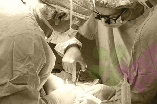


The name Bart syndrome entered the medical literature after being first reported by Dr. Bart in 1966, but its clinical classification remains a subject of debate. It differs from Bartter syndrome by only one letter, but the latter is a renal tubular genetic disorder involving electrolyte metabolism abnormalities, while Bart syndrome is entirely different. The core manifestation of Bart syndrome is localized skin loss present at birth, often accompanied by skin blisters and nail abnormalities. The academic community currently tends to view it as a clinical manifestation of epidermolysis bullosa rather than an independent disease entity.
Why is skin loss sometimes observed at birth?
At birth, infants with Bart syndrome typically present with well-defined skin defects on the legs, dorsum of the feet, or trunk, with a reddish, moist granulation tissue base. Sometimes the defect extends to the vicinity of the knee joint. This skin loss is not caused by trauma, but rather by increased fragility of the skin structure during fetal development, leading to separation of the epidermis from the dermis even under slight friction or pressure in utero. Bart syndrome blisters can appear on multiple sites, including the trunk, buttocks, elbows, and scalp. Rupture of the blisters forms erosions, with a slow healing process. Pathological analysis of skin biopsies indicates that Bart syndrome can correspond to any of the three main subtypes of epidermolysis bullosa: simple, junctional, or dystrophic, with the dystrophic subtype being more common. This subtype is characterized by skin cleavage occurring in the superficial dermis beneath the basement membrane.
In terms of familial inheritance, Bart syndrome typically follows an autosomal dominant inheritance pattern, affecting multiple family members. Studies show that in the originally reported Bart family, the gene mutation was located on the short arm of chromosome 3, specifically involving the COL7A1 gene encoding type VII collagen. Type VII collagen is a core component of anchoring fibers, responsible for firmly anchoring the epidermal basement membrane to the underlying dermis. Abnormalities in this protein structure significantly reduce the skin's tolerance to minor mechanical forces, leading to blisters and skin loss.
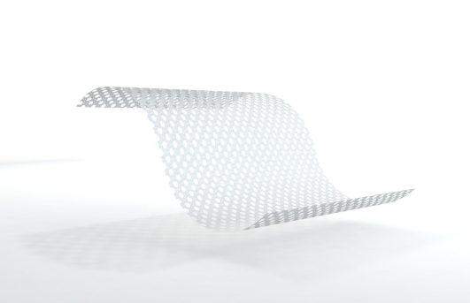
The core of Bart syndrome treatment lies in preventing skin damage and wound infection. Neonatal management is particularly critical, as extensive skin loss can lead to fluid loss, impaired thermoregulation, and protein depletion, which can be life-threatening in severe cases. Daily care requires covering exposed areas with non-adhesive dressings, avoiding direct application of tape to the skin. Clothing should be made of soft materials with seams facing outwards. For existing erosions or superficial ulcers, silicone gel foam dressings can be used. The silicone gel contact layer does not adhere to the moist wound, preventing the tearing of newly formed epidermal tissue during dressing changes. The foam layer absorbs small amounts of exudate and cushions external friction, a significant difference from ordinary dry gauze. Blisters must be managed by a professional. A sterile needle should be used to puncture the blister from the side to drain the fluid, leaving the blister top as a biological dressing to cover the wound; the blister top should not be directly torn off. Involvement of the oral or esophageal mucosa may cause feeding difficulties, requiring adjustments to food texture or the use of special feeding utensils. Perianal blisters may cause painful defecation, necessitating the use of stool softeners. As children grow, recurrent blisters and scarring at the joints can lead to finger and toe fusion or joint contractures, requiring long-term follow-up intervention from rehabilitation and orthopedic surgeons. For family members, if the pathogenic gene mutation is identified, prenatal diagnosis can be used for evaluation during subsequent pregnancies, including chorionic villus sampling or amniocentesis to obtain fetal cells for genetic testing. The prognosis of Bart syndrome is highly dependent on the specific subtype and the extent of organ involvement. Some severe subtypes of the borderline type have a high mortality rate in infancy, while milder subtypes of the malnourished type can survive into adulthood, but require long-term management of fragile skin and chronic wounds.
For more information on Innomed® Silicone Contact Layer, refer to the Previous Articles. If you have customized needs, you are welcome to contact us; You Wholeheartedly. At long-term medical, we transform this data by innovating and developing products that make life easier for those who need loving care.
Editor: kiki Jia


