 English
 عربى
عربى Español
 русский
 中文简体
中文简体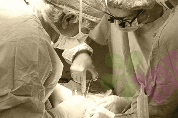


What is a giant pressure ulcer of the hip ?
Giant pressure ulcers of the hip typically refer to severe pressure ulcers occurring in the hip bone region, exceeding five centimeters in diameter or extending into the bone fascia. These wounds often develop due to prolonged bed rest or wheelchair use, leading to ischemia and necrosis of local tissues caused by continuous pressure. Giant pressure ulcers not only present as full-thickness skin defects but are often accompanied by exposure or necrosis of deeper tissues such as fat, muscle, and even bone, and may be accompanied by infection, exudation, and foul odor. Managing such wounds is a challenging task for both patients and healthcare professionals, as the repair process is complex, lengthy, and highly prone to recurrence.
Why is the first phase of restoration so difficult ?
The primary wound area of a large hip pressure ulcer typically has poor blood supply and lacks adequate soft tissue coverage, especially where bone or tendons are exposed. Secondly, the wound often contains undermining or sinus tracts, making complete removal of internal necrotic tissue difficult. Thirdly, the location is close to the ischial tuberosity, and patients are prone to pressure and shearing forces when sitting, standing, or turning over, affecting healing. Finally, patients often have underlying conditions such as malnutrition, diabetes, or neurological dysfunction, further weakening tissue repair capabilities. Therefore, simple suturing often fails, requiring more systematic and refined surgical repair strategies.
How to perform thorough and effective wound cleaning ?
All devitalized and necrotic soft tissue must be thoroughly removed, including dark-colored, non-contractile muscle and yellowish, softened adipose tissue. The bone surface must be cleaned with a bone chisel or burr until fresh, oozing bone cortex is visible. Debridement should extend to the edges of healthy tissue to ensure good blood supply to the wound base. Repeated irrigation and the use of disinfectant solutions such as hydrogen peroxide and povidone-iodine are necessary to reduce bacterial load. For severely infected wounds, multiple debridements may be required, with repair performed only after the infection is controlled.
How to design a flap to cover a large defect ?
Due to the large and deep wound, direct suturing would result in excessive tension and a high risk of dehiscence. Therefore, flap transfer is essential to provide adequate and well-vascularized tissue coverage. Commonly used and reliable flaps for the hip region include the gluteus maximus myocutaneous flap, the posterior femoral cutaneous nerve-vascular flap, the gracilis myocutaneous flap, or the tensor fasciae latae myocutaneous flap. Among these, the gluteus maximus myocutaneous flap is the preferred choice for repairing large defects in the hip and ischial tuberosity region due to its stable blood supply, ample tissue volume, and large rotational radius. The flap design must be larger than the wound surface to ensure tension-free suturing, and the flap pedicle should be protected from pressure or torsion to guarantee unobstructed blood flow.
How to ensure postoperative healing and prevent recurrence ?
A successful surgery is only the beginning; meticulous postoperative care is crucial. Patients must absolutely avoid pressure on the skin flap area, use a specially designed pressure-reducing mattress, and adhere to a strict turning schedule. Nutritional support must be provided, ensuring adequate calories, protein, vitamins, and micronutrients to promote tissue growth. Wound drainage tubes must be kept patent, and the skin flap's color, temperature, and swelling must be closely monitored for any vascular crisis. Healthcare professionals must instruct patients and their families on long-term pressure management, including proper posture, using pressure-reducing cushions, and daily skin checks. Even after the wound has healed, lifelong preventative measures are necessary, as the area with the previous pressure sore will always remain a vulnerable point.
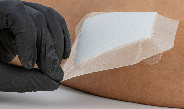
For more information on Innomed®Silicone Foam Dressing, Refer to the Previous Articles. If you have customized needs, you are welcome to contact us; You Wholeheartedly. At longterm medical, we transform this data by Innovating and Developing Products that Make Life easier for those who need loving care.
Editor: kiki Jia


