 English
 عربى
عربى Español
 русский
 中文简体
中文简体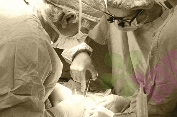


For those caring for wounds at home, the most pressing and anxiety-inducing question is whether the wound is healing. Looking at the wound under the dressing, the daily changes may seem subtle, sometimes even mistaking normal healing for signs of deterioration, leading to repeated changes in care and disrupting the recovery process. To accurately determine if a wound is healing, a set of workable observation criteria needs to be established, comprehensively assessing factors such as color, exudate, odor, pain, and the condition of the edges, rather than drawing conclusions based on a single momentary feeling.
Is it good or bad that the wound's color changes from black to red?
Changes in wound color are the most direct signal of healing. In a properly managed moist healing environment, a healing wound will show a clear evolutionary trajectory at its base. If initially covered with a black or brownish scab, improvement is indicated by the edges of the scab beginning to lift, revealing fresh tissue underneath, rather than the scab expanding or the surrounding skin becoming more red. If there was originally yellow necrotic tissue attached, improvement is shown by the gradual reduction of the necrotic area, replaced by small, bright red granulation tissue. This newly formed granulation tissue has a fine granular surface and bleeds easily upon touch, proving blood revascularization and tissue filling. As the wound progresses, the granulation tissue gradually becomes less bright red, turning pale pink and becoming smoother, indicating decreased blood vessel density and tissue maturation. It is important to distinguish between normal granulation tissue and edematous granulation tissue. The former is rosy and firm, while the latter is dark red and shiny, even protruding above the wound surface; the latter indicates hindered healing rather than improvement. Therefore, in summary, a change in wound color from dark to light, from dull to bright, and from disordered to uniform indicates a positive change.
Does low oozing indicate that healing is in progress?
Dynamic monitoring of exudate volume is more valuable for diagnosis than a single observation. In a wound healing smoothly, the amount of exudate will show a continuous decreasing trend. For example, initially, dressings may need to be changed daily for the first two days, but after a week, they may only need to be changed every two days without seepage. This is a typical improvement curve. However, a decrease in exudate should not be viewed in isolation; it needs to be assessed in conjunction with the characteristics of the exudate. Normally, the exudate during the healing process should be pale yellow, clear, or slightly cloudy, with a faint, normal bloody odor. This serous exudate contains growth factors and immune cells, which are the raw materials for healing. If the exudate, although reduced in volume, becomes viscous, yellowish-green, or even brown, or if a foul odor is present when the dressing is removed, this should not be considered a sign of improvement. It may indicate infection causing local pus concentration and poor drainage. Another easily misunderstood situation is when the wound surface appears dry, but this is actually dehydrated scabs formed from exposure to air. These scabs often conceal a stagnant, atrophied wound, not true healing. Therefore, when observing exudate, it is crucial to grasp the balance between quantity and quality; a decrease in quantity and clarity is reliable.
What changes at the edges of a wound should reassure you?
The condition of the wound edges directly reflects the epithelialization process, which is the process of the outermost layer of skin regrowing. In a healing wound, the edges will change from indistinct to well-defined, and a thin, pinkish-white, translucent membrane will appear, creeping from the edges towards the center. This is the result of keratinocyte migration and proliferation, a sign that healing has entered its final stage. Simultaneously, the previously red, swollen, and hardened edges will gradually soften, changing color from dark red to a tone closer to the surrounding normal skin. When touched lightly, the temperature will be the same as the surrounding area, no longer hot. If the wound is small, the edges will appear to close inward within a few weeks, and the overall area will shrink; this process is slow but consistently visible. Another positive sign is that the inward or everted edges of the wound begin to improve, indicating that epithelial growth is no longer hindered. Conversely, if the edges remain sharp and angular, the surrounding red halo expands and becomes raised, and the skin feels hard, it suggests ongoing inflammation and should not be considered an improvement. Observing these edge changes requires ample natural light, and taking photos for comparison each time will provide clearer results.
Is the wound healing as the pain gradually subsides?
Pain is a subjective experience with significant individual differences, but it still has reference value as an indicator. Generally, as the inflammatory response subsides and new tissue covers nerve endings, the resting pain of the wound will be relieved first, meaning there will be no more throbbing or burning sensation when not touched or moved. The pain during dressing changes will also gradually decrease because fewer nerve endings are exposed. However, if the nature of the pain changes, caution is needed. For example, if the dull pain suddenly becomes a sharp, needle-like pain that intensifies intermittently, it may not be an improvement but rather neuropathic pain or a worsening of local infection. In addition, in some ischemic wounds, as blood supply improves during the healing process, previously numb and painless areas may regain sensation, which is actually a good sign of nerve function recovery. Therefore, the significance of pain cannot be judged in a one-size-fits-all manner; it must be considered within the context of the overall changes in the wound. When pain reduction occurs simultaneously with the wound turning red, exudation decreasing, and the edges contracting, it can be confirmed that the wound is improving.
Simple and easy methods for home observation :
Relying solely on memory to compare wound changes can easily lead to inaccuracies; establishing a simple timeline record is essential. Each time the dressing is changed, take a photo with your phone under the same lighting and angle, placing a ruler nearby for reference. After a week of continuous recording, review the photos to clearly see changes in area and color. Simultaneously, briefly jot down the amount of exudate, odor, and pain score. The score can be quantified from zero to ten, where zero indicates no pain at all, and ten indicates unbearable pain. If the pain score consistently decreases by two to three points over several days, it represents clear progress. If any of the following conditions are observed—failure to improve or even worsening after two consecutive dressing changes—prompt professional evaluation is necessary: the wound area expands instead of shrinking; granulation tissue changes from red to dark purple or pale; a sudden increase in exudate; surrounding redness and swelling exceeding one centimeter with fever; or pain changes from tolerable to persistent throbbing. Mastering these judgment criteria will significantly reduce anxiety associated with home care, replacing it with a sense of reassurance based on evidence.
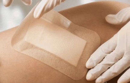
For more information on Innomed® Silicone Contact Layer, refer to the Previous Articles. If you have customized needs, you are welcome to contact us; You Wholeheartedly. At long-term medical, we transform this data by innovating and developing products that make life easier for those who need loving care.
Editor: kiki Jia


