 English
 عربى
عربى Español
 русский
 中文简体
中文简体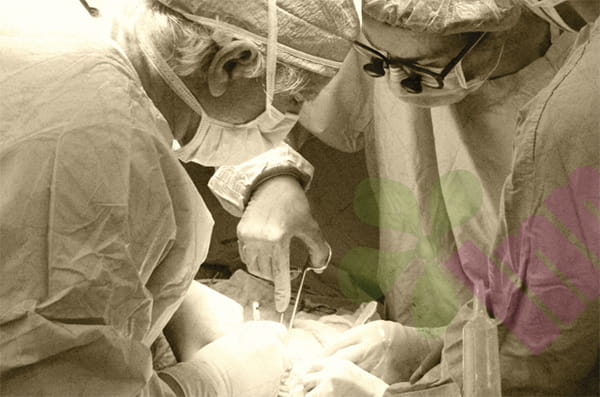


Hand-foot syndrome, also known as palmoplantar erythema with loss of sensation, is a skin toxicity reaction caused by certain chemotherapy drugs or targeted therapies. Its pathological mechanism is mainly related to the accumulation of drugs and their metabolites in the capillary-rich tissues of the hands and feet. These high concentrations of toxic substances directly damage keratinocytes, inducing an inflammatory response. Clinically, it progresses from prodromal palmoplantar sensory abnormalities and erythema to dryness, desquamation, blisters, and ulcers. From a wound care perspective, when the integrity of the skin is compromised due to ruptured blisters, cracking, or ulceration, hand-foot syndrome is no longer merely an adverse drug reaction but constitutes an acute wound requiring professional intervention. These wounds are characterized by indistinct borders, fragile surrounding skin, and patients often have a background of immunosuppression; therefore, their treatment principles differ significantly from those for routine mechanical injuries.
How can skin care help prevent wounds?
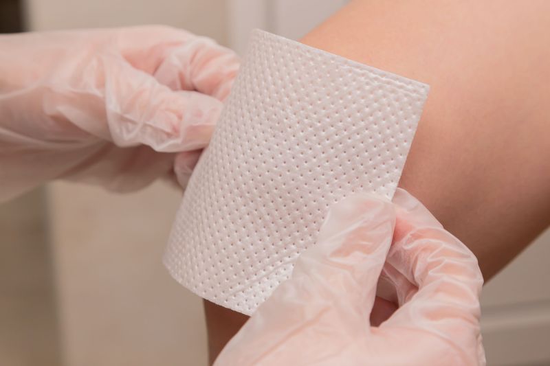
Effective wound prevention begins with proactively maintaining the skin barrier function. The goal of this stage is to prevent the progression from the erythematous stage to the lesion stage. The core measures are strengthening the hydration of the stratum corneum and reducing local friction. It is recommended to use fragrance-free, preservative-free urea ointment or ceramide-based moisturizing barrier repair cream, applied multiple times daily in a thick layer, especially effective when applied within three minutes after bathing when the skin is still slightly damp. Urea has a gentle keratolytic effect and powerful moisturizing properties, increasing the water content of the stratum corneum, thereby improving skin elasticity and resisting mechanical stress damage. Simultaneously, patients should be instructed to systematically avoid risk factors, including wearing seamless, absorbent cotton socks and loose-fitting, soft-soled shoes, and avoiding prolonged walking, standing, and repetitive hand grasping movements. Physical barriers are also crucial; cotton-lined rubber gloves should be worn when doing housework, and contact with hot water and strong detergents should be avoided. Through this strict skin management program, the incidence of skin damage can be significantly reduced, making it the most cost-effective link in the entire management chain.
How should we intervene in accordance with wound care principles?
When skin damage has progressed to the blistering or erosion stage, the treatment principle must immediately shift to standard wound care procedures. The core principles are protecting the wound, controlling exudation, and preventing infection. For tension blisters less than one centimeter in diameter located in non-weight-bearing areas, the principle of preserving the intact blister skin should be followed, as it can act as a natural biological dressing to isolate external bacteria. After cleaning with saline solution, a sterile foam dressing can be applied to absorb any small amount of exudate and provide cushioning. For blisters larger than one centimeter in diameter, or located in high-friction areas such as the heel or knuckles, the blister skin is highly susceptible to accidental rupture. It is recommended to aspirate the blister fluid with a fine needle under strict aseptic conditions, but it is essential to preserve the blister skin intact, adhere it to the wound, and then cover it with a dressing. For erosions that have ruptured and exposed the dermis, gently rinse with room temperature saline solution to remove residual blister fluid and devitalized tissue. The choice of dressing depends on the amount of exudate: for small amounts of exudate, silicone foam dressings are the first choice, as they provide a moist healing environment and are painless to change without damaging the surrounding delicate skin; for moderate to large amounts of exudate, alginate fiber dressings or hydrophilic fiber dressings with water-locking properties can be used as the inner layer to absorb the exudate, with the outer layer secured with foam dressings or gauze. Strong adhesive tapes should never be used, as they may cause new skin avulsion injuries when removed.
How to manage and control the risk of infection?
Deep fissures and ulcers are the most severe skin manifestations of hand-foot syndrome, causing intense pain and carrying a very high risk of infection. These wounds are often accompanied by significant inflammation and tissue loss. The cleaning phase should be more meticulous; after rinsing with saline solution, gently roll sterile cotton swabs to remove fibrinous necrotic tissue. This process must be extremely gentle to avoid pain and bleeding. For dressings, for dry fissures with a hard scab at the base, hydrogel dressings can be considered for autolytic debridement, softening necrotic tissue by providing moisture and effectively relieving pain. For ulcers with significant exudation and potential signs of infection, such as increased redness, swelling, heat, pain, or purulent discharge, medical-grade antibacterial dressings, such as foam or fiber dressings containing silver ions or polyhexamethylene biguanide, should be used under the guidance of a physician, based on microbial culture results. These dressings can continuously release broad-spectrum antibacterial components into the wound, effectively controlling the local bioburden. Given that chemotherapy patients may have decreased neutrophils, any signs of local infection must be closely monitored, as they can rapidly progress to cellulitis or even sepsis. This stage requires daily wound assessment and close communication with oncologists to determine whether the chemotherapy regimen needs to be adjusted or medication needs to be suspended.
How to manage persistent peeling and cracking in the later stages of wound healing?
After acute inflammation subsides and the wound has largely healed, the skin enters a prolonged repair period, characterized by persistent hyperkeratosis, extensive scaling, and cracking due to decreased elasticity. The focus of treatment during this stage shifts from wound closure to the reconstruction of the skin barrier and the restoration of its function. Continued and intensified use of potent moisturizing creams containing urea or lactic acid is recommended. For abnormally thickened stratum corneum, high-concentration urea ointment can be used intermittently under a doctor's guidance to gently exfoliate. For small cracks that reappear due to dryness, a liquid skin protectant can be applied to form a transparent, breathable protective film, effectively blocking irritation and preventing the cracks from deepening. This entire process may take weeks or even months until the skin recovers to near-normal thickness and elasticity; long-term maintenance is crucial. For more information on Innomed®Silicone Contact Layer, refer to the Previous Articles. If you have customized needs, you are welcome to contact us; You Wholeheartedly. At long-term medical, we transform this data by innovating and developing products that make life easier for those who need loving care.
Editor: kiki Jia


