 English
 عربى
عربى Español
 русский
 中文简体
中文简体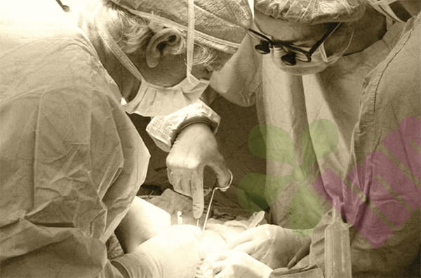


Duke University and Nokia Bell Labs have jointly developed a new platform that can objectively and non-invasively monitor the healing process and optimize the design of biomaterials.
Clinical monitoring of the wound healing process has long faced challenges. Regardless of the size of the wound, traditional methods such as biopsies damage tissue and are unsuitable for repeated testing; while most high-precision imaging devices are bulky, expensive, and often occupied by more urgent diagnostic and treatment needs. Currently, doctors mostly rely on visual observation or simple measurement of wound size changes.
Biomedical engineers at Duke University, in collaboration with Nokia Bell Labs, have developed a novel imaging system that combines optical coherence tomography (OCT) with artificial intelligence (AI) to accurately and objectively assess wound healing processes. The study also found that a hydrogel used to promote healing exhibits better healing effects when its mechanical properties are stiffer. These findings were published online in the journal *Cell Biomaterials* on March 20.
“Wound healing is a complex process, and surface observations don’t always reflect the true internal situation,” said Sharon Gerecht, Duke University’s Paul M. Gross Distinguished Professor of Biomedical Engineering and the lead researcher. “For over a decade, my lab has been working on hydrogel therapies. This collaboration with Nokia Bell Labs allows us to combine advanced optical imaging with AI to gain an unprecedented understanding of how biomaterials induce healing deep within tissues.”
OCT technology is best known for its application in ophthalmology, where it provides three-dimensional images of the fundus for diagnosing retinal diseases. In this study, researchers applied its depth-resolution imaging capabilities to wound monitoring, using light energy to non-invasively observe subcutaneous tissue structures and blood flow dynamics.
However, imaging alone is far from sufficient to transform massive amounts of OCT data into meaningful biological information. In a collaborative project spanning several years, Nokia Bell Labs developed a customized OCT system and trained an AI analysis model based on datasets collected by Grecht's lab. This OCT-AI platform can automatically quantify the evolution of tissue structure and vascular dynamics over time, achieving objective healing assessments that surpass traditional visualization methods.
To validate the platform's effectiveness, the research team applied it to mouse wounds treated with a hydrogel developed by Grecht's lab and compared the mechanical properties of softer and harder hydrogels. During a two-week observation period, the platform demonstrated in detail how granulation tissue—the glassy tissue that initially fills the wound—gradually fills the gaps and matures. Data showed that the harder hydrogel promoted the formation of more initial granulation tissue in a shorter time and accelerated its transition to complete regenerated tissue.
“With this technology, we can monitor blood flow near the wound and understand structural and vascular changes in real time,” said Zhiyan Song, a postdoctoral researcher in the Grecht Lab and co-first author of the paper. “Artificial intelligence helps us quantitatively track these changes and obtain more objective results without having to manually analyze images.”
The research team plans to further advance the clinical translation of this platform. While OCT-AI has proven effective in its current relatively simple scenarios, significant work remains to be done to expand its application from monitoring healing processes to predicting a wider range of disease states. Grecht's team is seeking research funding to refine the system, enabling it to predict the healing of chronic wounds in diabetic patients.
This research was supported by the P30 Cancer Center Support Fund (P30 CA014236), the American Heart Association, the Duke Regenerative Center (DRC), Duke Science and Technology (DST), and Nokia Bell Labs.


.jpg.png "Ostomy Pouch 210ML")


.jpg.png "Colostomy Pouch 350ML Closed")


